IPLab:Lab 4:Septic Emboli: Difference between revisions
Jump to navigation
Jump to search
Seung Park (talk | contribs) |
Seung Park (talk | contribs) No edit summary |
||
| Line 18: | Line 18: | ||
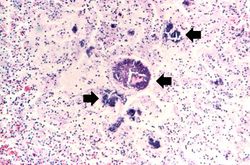
File:IPLab4SepticEmboli8.jpg|This is a high-powered photomicrograph of a myocardial abscess stained with a special tissue Gram stain (Brown & Brenn) to illustrate the colonies of bacteria in this myocardial tissue (arrows). | File:IPLab4SepticEmboli8.jpg|This is a high-powered photomicrograph of a myocardial abscess stained with a special tissue Gram stain (Brown & Brenn) to illustrate the colonies of bacteria in this myocardial tissue (arrows). | ||
</gallery> | </gallery> | ||
== Virtual Microscopy == | |||
<peir-vm>IPLab4SepticEmboli</peir-vm> | |||
== Study Questions == | == Study Questions == | ||
Revision as of 16:12, 3 January 2014
Clinical Summary
This 4-year-old female sustained second and third degree burns involving approximately forty percent of the body surface. Subsequently, she developed septicemia secondary to skin infection and died in septic shock on the 10th hospital day. Antemortem blood cultures were positive for Enterobacter aerogenes and Staphylococcus aureus.
Autopsy Findings
Postmortem findings included (1) multiple abscesses diffusely distributed throughout the parenchyma of the lung and heart, (2) lobular pneumonia, and (3) visceral congestion.
Images
-
 This gross photograph of lung demonstrates microabscesses due to septic embolization. Note the small 2 to 3-mm lesions scattered throughout this lung tissue (arrows).
 -
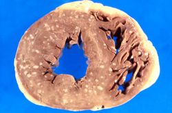 This is a gross photograph of myocardium with multiple embolic lesions scattered throughout the left and right ventricles.
 -
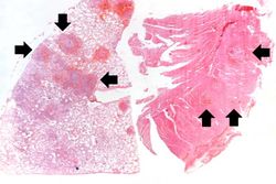 This low-power photomicrograph shows a section of lung on the left and myocardium on the right. Both pieces of tissue have multiple embolic lesions seen as blue staining areas with massive infiltration of inflammatory cells (arrows).
 -
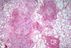
-
-

-
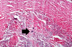 This is a higher-power photomicrograph of the abscess within the myocardium illustrating colonies of bacteria as the dark blue-staining material (arrow).
 -
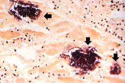 This is a high-powered photomicrograph of a myocardial abscess stained with a special tissue Gram stain (Brown & Brenn) to illustrate the colonies of bacteria in this myocardial tissue (arrows).


Virtual Microscopy
<peir-vm>IPLab4SepticEmboli</peir-vm>
Study Questions
- <spoiler text="What is the most common cause of septicemia in burn patients?">Pseudomona aeruginosa.</spoiler>
- <spoiler text="What factors dictate the location of the abscesses formed by septic emboli?">Distribution of blood flow downstream from the source of septic emboli.</spoiler>
Additional Resources
Reference
- eMedicine Medical Library: Septic Shock
- eMedicine Medical Library: Bacterial Sepsis
- eMedicine Medical Library: Multisystem Organ Failure of Sepsis
- Merck Manual: Sepsis and Septic Shock
- Merck Manual: Bacteremia
Journal Articles
- Hiorns MP, Screaton NJ, Müller NL. Acute lung disease in the immunocompromised host. Radiol Clin North Am 2001 Nov;39(6):1137-51, vi.
Images
- PEIR Digital Library: Vasculitis Due to Septic Embolus Images
- WebPath: Pulmonary Thromboembolus
- WebPath: Atherosclerosis and Thrombosis
Related IPLab Cases
| |||||