File:IPLab1KidneyInfarction5.jpg: Difference between revisions
Jump to navigation
Jump to search

Seung Park (talk | contribs) No edit summary |
Seung Park (talk | contribs) No edit summary |
||
| Line 1: | Line 1: | ||
This higher-power view of the infarct demonstrates retention of the tubular structure and cellular outlines. In the lower right-hand corner is a barely identifiable glomerulus (1). Note that, although the cellular architecture is retained, there are no nuclei within the renal tubular cells. The nuclei visible in this photomicrograph are the nuclei of inflammatory cells. | |||


Revision as of 11:18, 15 August 2013
This higher-power view of the infarct demonstrates retention of the tubular structure and cellular outlines. In the lower right-hand corner is a barely identifiable glomerulus (1). Note that, although the cellular architecture is retained, there are no nuclei within the renal tubular cells. The nuclei visible in this photomicrograph are the nuclei of inflammatory cells.
File history
Click on a date/time to view the file as it appeared at that time.
| Date/Time | Thumbnail | Dimensions | User | Comment | |
|---|---|---|---|---|---|
| current | 16:20, 27 June 2019 | 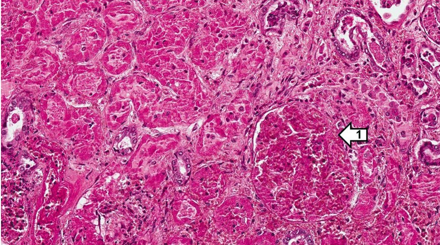 | 869 × 485 (242 KB) | Peter Anderson (talk | contribs) | |
| 10:12, 15 August 2013 | 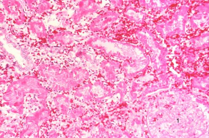 | 679 × 450 (64 KB) | Seung Park (talk | contribs) |
You cannot overwrite this file.
File usage
The following page uses this file:
