IPLab:Lab 4:Atheromatous Emboli: Difference between revisions
Jump to navigation
Jump to search
Seung Park (talk | contribs) Created page with "== Images == <gallery heights="250px" widths="250px"> File:IPlab4AtheromatousEmboli1.jpg|This is a gross photograph of the aorta from this patient opened lengthwise with the l..." |
Seung Park (talk | contribs) No edit summary |
||
| Line 1: | Line 1: | ||
== Images == | == Images == | ||
<gallery heights="250px" widths="250px"> | <gallery heights="250px" widths="250px"> | ||
File: | File:IPLab4AtheromatousEmboli1.jpg|This is a gross photograph of the aorta from this patient opened lengthwise with the luminal surface visible. Note the rough surface with ulcerations and adherent thrombotic material. There is a mild dilation (aneurysm) at the distal aorta just at the bifurcation with an accumulation of thrombus. | ||
File: | File:IPLab4AtheromatousEmboli2.jpg|This is a closer view of the luminal surface of the aorta from the previous image. The rough, ulcerated surface and the thrombotic material can be easily seen in this image. | ||
File: | File:IPLab4AtheromatousEmboli3.jpg|This is a low-power photomicrograph of kidney tissue. Several blood vessels can be identified at the corticomedullary junction (arrows). | ||
File: | File:IPLab4AtheromatousEmboli4.jpg|This higher-power photomicrograph of one of the arcuate arteries reveals a cholesterol embolus. Note the needle-shaped space (arrow) within the lumen of this artery (arrow) which represents the space occupied by the cholesterol crystal that was dissolved away during histologic processing. | ||
File: | File:IPLab4AtheromatousEmboli5.jpg|This is another view of this vessel with an atherosclerotic embolus. Note the cholesterol clefts (1) and thrombotic material (2) that occlude this artery. | ||
File: | File:IPLab4AtheromatousEmboli6.jpg|A mesenteric artery also had an atherosclerotic embolus. Again note the cholesterol clefts and thrombotic material that occlude this artery. | ||
</gallery> | </gallery> | ||
Revision as of 12:02, 19 August 2013
Images
-
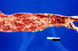 This is a gross photograph of the aorta from this patient opened lengthwise with the luminal surface visible. Note the rough surface with ulcerations and adherent thrombotic material. There is a mild dilation (aneurysm) at the distal aorta just at the bifurcation with an accumulation of thrombus.
 -
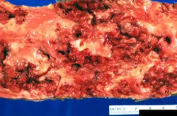 This is a closer view of the luminal surface of the aorta from the previous image. The rough, ulcerated surface and the thrombotic material can be easily seen in this image.
 -
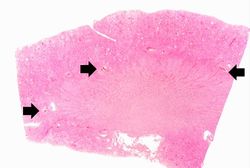 This is a low-power photomicrograph of kidney tissue. Several blood vessels can be identified at the corticomedullary junction (arrows).
 -
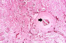 This higher-power photomicrograph of one of the arcuate arteries reveals a cholesterol embolus. Note the needle-shaped space (arrow) within the lumen of this artery (arrow) which represents the space occupied by the cholesterol crystal that was dissolved away during histologic processing.
 -
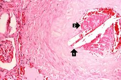 This is another view of this vessel with an atherosclerotic embolus. Note the cholesterol clefts (1) and thrombotic material (2) that occlude this artery.
 -
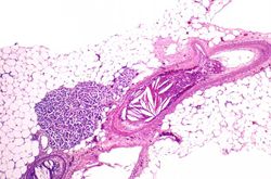 A mesenteric artery also had an atherosclerotic embolus. Again note the cholesterol clefts and thrombotic material that occlude this artery.


| |||||