File:IPLab2Hypertrophy4.jpg: Difference between revisions
Jump to navigation
Jump to search
Seung Park (talk | contribs) Normal myocardium (left) is compared to hypertrophied myocardium (right). This high power view demonstrates the large dark nuclei (arrow) found in hypertrophied cardiac muscle cells. Polyploidy is a common feature in cardiac hypertrophy. Also note the ... |
Peter Anderson uploaded a new version of "File:IPLab2Hypertrophy4.jpg" |
(No difference)
| |


Latest revision as of 17:27, 4 September 2013
Normal myocardium (left) is compared to hypertrophied myocardium (right). This high power view demonstrates the large dark nuclei (arrow) found in hypertrophied cardiac muscle cells. Polyploidy is a common feature in cardiac hypertrophy. Also note the increased size (thickness) of the individual cardiac muscle cell on the right compared to normal cardiac myocytes (left).
File history
Click on a date/time to view the file as it appeared at that time.
| Date/Time | Thumbnail | Dimensions | User | Comment | |
|---|---|---|---|---|---|
| current | 17:27, 4 September 2013 | 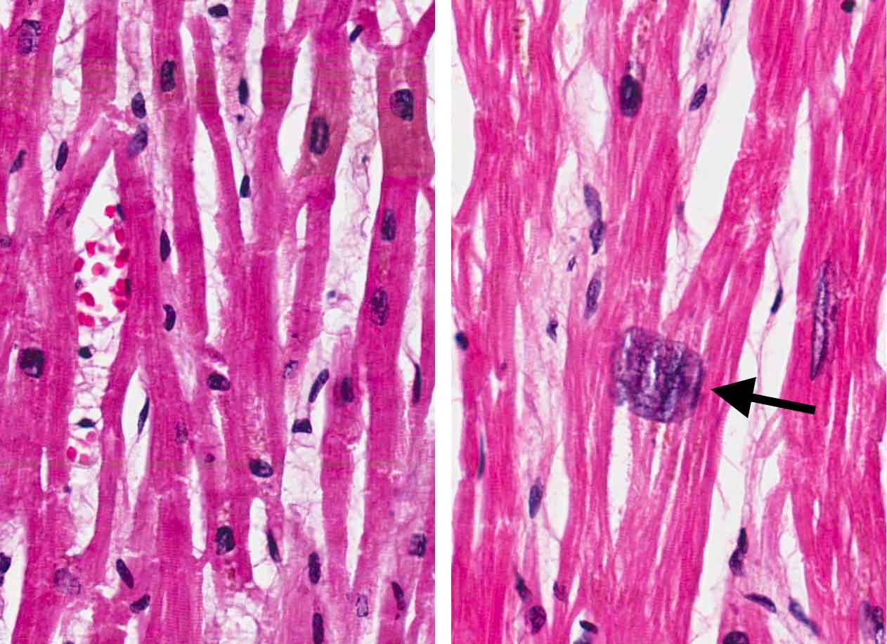 | 1,262 × 916 (125 KB) | Peter Anderson (talk | contribs) | |
| 18:29, 18 August 2013 | 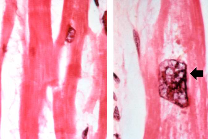 | 673 × 450 (30 KB) | Seung Park (talk | contribs) | Normal myocardium (left) is compared to hypertrophied myocardium (right). This high power view demonstrates the large dark nuclei (arrow) found in hypertrophied cardiac muscle cells. Polyploidy is a common feature in cardiac hypertrophy. Also note the ... |
You cannot overwrite this file.
File usage
The following page uses this file:
