File:IPLab1Tuberculosis4.jpg: Difference between revisions
Jump to navigation
Jump to search
Seung Park (talk | contribs) No edit summary |
Seung Park (talk | contribs) No edit summary |
||
| Line 1: | Line 1: | ||
This is a low-power photomicrograph of a histology section from the lung of this patient with a chronic history of respiratory disease. Note the multiple eosinophilic nodules (arrows) seen at low power in this section. Other areas of the lung are relatively normal and several bronchi and large vessels can be seen at this low power. The pleural surface of the lung is at the left and the remaining edges are cut edges of the tissue block. | |||


Latest revision as of 21:52, 15 August 2013
This is a low-power photomicrograph of a histology section from the lung of this patient with a chronic history of respiratory disease. Note the multiple eosinophilic nodules (arrows) seen at low power in this section. Other areas of the lung are relatively normal and several bronchi and large vessels can be seen at this low power. The pleural surface of the lung is at the left and the remaining edges are cut edges of the tissue block.
File history
Click on a date/time to view the file as it appeared at that time.
| Date/Time | Thumbnail | Dimensions | User | Comment | |
|---|---|---|---|---|---|
| current | 21:50, 15 August 2013 | 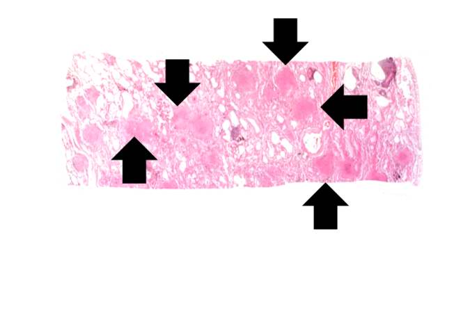 | 675 × 450 (21 KB) | Seung Park (talk | contribs) |
You cannot overwrite this file.
File usage
There are no pages that use this file.
