IPLab:Lab 4:Atheromatous Emboli: Difference between revisions
Jump to navigation
Jump to search
Seung Park (talk | contribs) No edit summary |
|||
| (4 intermediate revisions by 2 users not shown) | |||
| Line 1: | Line 1: | ||
== Clinical Summary == | == Clinical Summary == | ||
This 71-year-old male was admitted to the hospital with dry gangrene of the left great toe. Subsequently, he was found to have a poorly differentiated adenocarcinoma of the left upper lobe of the lung. He developed a perforation of a duodenal ulcer with generalized peritonitis and died five days after admission. | |||
Gross examination of the aorta revealed severe atherosclerosis with extensive mural thrombus formation. Recent infarcts were noted in the spleen and in the left kidney. An old posterior myocardial infarct was noted. The left iliac artery also appeared to be almost totally occluded. A whitish-tan tumor was noted in the apical portion of the left upper lobe of the lung with metastases in the carinal and hilar lymph nodes. | |||
Gross examination of the aorta revealed severe atherosclerosis with extensive mural thrombus formation. Recent infarcts were noted in the spleen and in the left kidney. An old posterior myocardial infarct was noted. The left iliac artery also appeared to be almost totally occluded. A whitish-tan tumor was noted in the apical portion of the left upper lobe of the lung with metastases in the carinal and hilar lymph nodes. | |||
== Images == | == Images == | ||
| Line 16: | Line 13: | ||
File:IPLab4AtheromatousEmboli6.jpg|A mesenteric artery also had an atherosclerotic embolus. Again note the cholesterol clefts and thrombotic material that occlude this artery. | File:IPLab4AtheromatousEmboli6.jpg|A mesenteric artery also had an atherosclerotic embolus. Again note the cholesterol clefts and thrombotic material that occlude this artery. | ||
</gallery> | </gallery> | ||
== Virtual Microscopy == | |||
=== Kidney: Atheromatous Emboli === | |||
<peir-vm>IPLab4AtheromatousEmboli</peir-vm> | |||
=== Normal Kidney === | |||
<peir-vm>UAB-Histology-00114</peir-vm> | |||
== Study Questions == | == Study Questions == | ||
| Line 32: | Line 36: | ||
=== Images === | === Images === | ||
* [ | * [{{SERVER}}/library/index.php?/tags/52-kidney PEIR Digital Library: Kidney Images] | ||
* [http://library.med.utah.edu/WebPath/ATHHTML/ATHIDX.html WebPath: Atherosclerosis and Thrombosis] | * [http://library.med.utah.edu/WebPath/ATHHTML/ATHIDX.html WebPath: Atherosclerosis and Thrombosis] | ||
* [http://library.med.utah.edu/WebPath/CVHTML/CVIDX.html WebPath: Cardiovascular Pathology] | * [http://library.med.utah.edu/WebPath/CVHTML/CVIDX.html WebPath: Cardiovascular Pathology] | ||
Latest revision as of 21:01, 23 June 2020
Clinical Summary
This 71-year-old male was admitted to the hospital with dry gangrene of the left great toe. Subsequently, he was found to have a poorly differentiated adenocarcinoma of the left upper lobe of the lung. He developed a perforation of a duodenal ulcer with generalized peritonitis and died five days after admission.
Gross examination of the aorta revealed severe atherosclerosis with extensive mural thrombus formation. Recent infarcts were noted in the spleen and in the left kidney. An old posterior myocardial infarct was noted. The left iliac artery also appeared to be almost totally occluded. A whitish-tan tumor was noted in the apical portion of the left upper lobe of the lung with metastases in the carinal and hilar lymph nodes.
Images
-
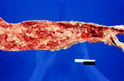 This is a gross photograph of the aorta from this patient opened lengthwise with the luminal surface visible. Note the rough surface with ulcerations and adherent thrombotic material. There is a mild dilation (aneurysm) at the distal aorta just at the bifurcation with an accumulation of thrombus.
 -
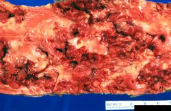 This is a closer view of the luminal surface of the aorta from the previous image. The rough, ulcerated surface and the thrombotic material can be easily seen in this image.
 -
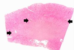 This is a low-power photomicrograph of kidney tissue. Several blood vessels can be identified at the corticomedullary junction (arrows).
 -
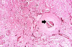 This higher-power photomicrograph of one of the arcuate arteries reveals a cholesterol embolus. Note the needle-shaped space (arrow) within the lumen of this artery (arrow) which represents the space occupied by the cholesterol crystal that was dissolved away during histologic processing.
 -
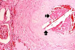 This is another view of this vessel with an atherosclerotic embolus. Note the cholesterol clefts (1) and thrombotic material (2) that occlude this artery.
 -
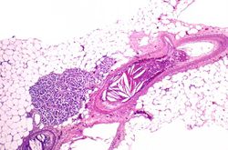 A mesenteric artery also had an atherosclerotic embolus. Again note the cholesterol clefts and thrombotic material that occlude this artery.


Virtual Microscopy
Kidney: Atheromatous Emboli
Normal Kidney
Study Questions
Additional Resources
Reference
- eMedicine Medical Library: Noncoronary Atherosclerosis
- eMedicine Medical Library: Cholesterol Embolism
- Merck Manual: Thrombotic Disorders
Journal Articles
- Scolari F, Ravani P. Atheroembolic renal disease. Lancet 2010 May 8;375(9726):1650-60.
Images
- PEIR Digital Library: Kidney Images
- WebPath: Atherosclerosis and Thrombosis
- WebPath: Cardiovascular Pathology
- WebPath: Renal Pathology
Related IPLab Cases
| |||||