Difference between revisions of "File:IPLab6TB4.jpg"
(This is a higher-power photomicrograph of a TB granuloma. The area of caseous necrosis is on the left side of the image, there are multinucleated giant cells and epithelioid macrophages throughout the remainder of the tissue.) |
(No difference)
|
Latest revision as of 20:11, 20 August 2013
This is a higher-power photomicrograph of a TB granuloma. The area of caseous necrosis is on the left side of the image, there are multinucleated giant cells and epithelioid macrophages throughout the remainder of the tissue.
Caseous means cheesy.
File history
Click on a date/time to view the file as it appeared at that time.
| Date/Time | Thumbnail | Dimensions | User | Comment | |
|---|---|---|---|---|---|
| current | 20:11, 20 August 2013 | 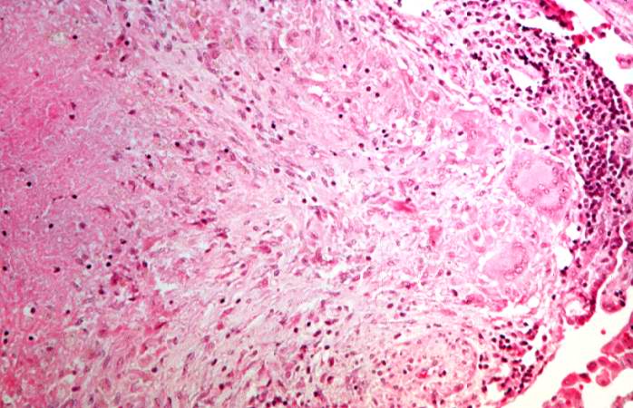 | 698 × 450 (68 KB) | Peter Anderson (talk | contribs) | This is a higher-power photomicrograph of a TB granuloma. The area of caseous necrosis is on the left side of the image, there are multinucleated giant cells and epithelioid macrophages throughout the remainder of the tissue. |
- You cannot overwrite this file.
File usage
The following page links to this file: