Difference between revisions of "File:IPLab5Gout5.jpg"
(This is a higher-power photomicrograph of the edge of the tophus. Most of the urate crystals dissolve away during processing. The inflammatory cells at the edge of these foci are clearly visible (arrow).) |
(No difference)
|
Latest revision as of 15:19, 20 August 2013
This is a higher-power photomicrograph of the edge of the tophus. Most of the urate crystals dissolve away during processing. The inflammatory cells at the edge of these foci are clearly visible (arrow).
A tophus is a chalky accumulation of urate crystals found in the tissue surrounding a joint.
File history
Click on a date/time to view the file as it appeared at that time.
| Date/Time | Thumbnail | Dimensions | User | Comment | |
|---|---|---|---|---|---|
| current | 15:19, 20 August 2013 | 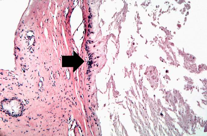 | 688 × 450 (60 KB) | Peter Anderson (talk | contribs) | This is a higher-power photomicrograph of the edge of the tophus. Most of the urate crystals dissolve away during processing. The inflammatory cells at the edge of these foci are clearly visible (arrow). |
- You cannot overwrite this file.
File usage
The following page links to this file: