Difference between revisions of "File:IPLab5PolycysticKidney4.jpg"
(This is a low-power photomicrograph of an H&E-stained section from this polycystic kidney. Note the large cystic structures (1), the few residual glomeruli (2), and the fibrous connective tissue throughout this section.) |
(No difference)
|
Latest revision as of 17:51, 19 August 2013
This is a low-power photomicrograph of an H&E-stained section from this polycystic kidney. Note the large cystic structures (1), the few residual glomeruli (2), and the fibrous connective tissue throughout this section.
File history
Click on a date/time to view the file as it appeared at that time.
| Date/Time | Thumbnail | Dimensions | User | Comment | |
|---|---|---|---|---|---|
| current | 17:51, 19 August 2013 | 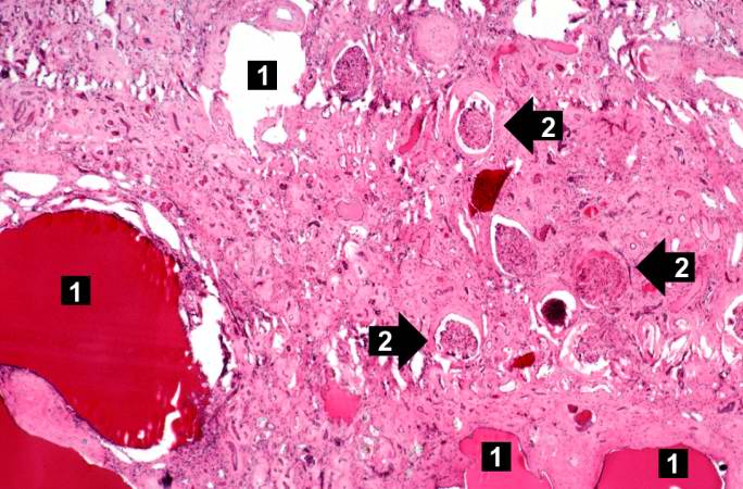 | 684 × 450 (69 KB) | Peter Anderson (talk | contribs) | This is a low-power photomicrograph of an H&E-stained section from this polycystic kidney. Note the large cystic structures (1), the few residual glomeruli (2), and the fibrous connective tissue throughout this section. |
- You cannot overwrite this file.
File usage
The following page links to this file: