Difference between revisions of "File:IPLab2Metaplasia1.jpg"
(This is a low-power photomicrograph showing the full cortical and medullary thickness of the kidney. Note that there is a dilated calyx containing some red blood cells in the center of the section (arrow). The cortex is markedly thin and has severe les...) |
(No difference)
|
Latest revision as of 15:39, 19 August 2013
This is a low-power photomicrograph showing the full cortical and medullary thickness of the kidney. Note that there is a dilated calyx containing some red blood cells in the center of the section (arrow). The cortex is markedly thin and has severe lesions of degeneration and atrophy, although these are hard to appreciate at this low magnification.
File history
Click on a date/time to view the file as it appeared at that time.
| Date/Time | Thumbnail | Dimensions | User | Comment | |
|---|---|---|---|---|---|
| current | 15:39, 19 August 2013 | 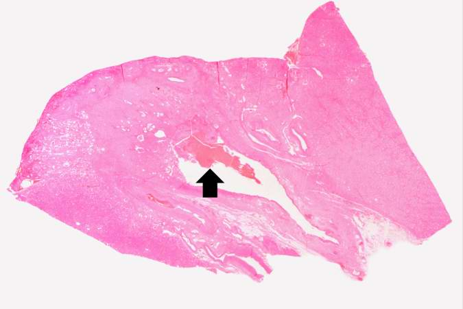 | 675 × 450 (26 KB) | Peter Anderson (talk | contribs) | This is a low-power photomicrograph showing the full cortical and medullary thickness of the kidney. Note that there is a dilated calyx containing some red blood cells in the center of the section (arrow). The cortex is markedly thin and has severe les... |
- You cannot overwrite this file.
File usage
There are no pages that link to this file.