IPLab:Lab 10:Blastomycosis: Difference between revisions
Jump to navigation
Jump to search
Seung Park (talk | contribs) |
|||
| (9 intermediate revisions by 2 users not shown) | |||
| Line 1: | Line 1: | ||
== Clinical Summary == | == Clinical Summary == | ||
About three weeks before his death, this 17-year-old white male developed a "chest cold" which gradually worsened. The patient was eventually admitted three days before his death. At that time, the patient was very dyspneic. Chest x-ray showed consolidation of the entire left lung. The initial impression by his care team was staphylococcal pneumonia. However, Blastomyces dermatitides was identified in stained smears of sputum the next day. In spite of appropriate antifungal therapy, the patient deteriorated rapidly and died. | |||
At autopsy the both lungs had areas of consolidation and necrosis. | |||
== Images == | == Images == | ||
| Line 20: | Line 17: | ||
File:IPLab10Blasto10.jpg|This is a very high-power photomicrograph showing Blastomyces organisms stained with PAS. Note the budding organism (arrow) and the underlying pyogranulomatous inflammatory reaction in the background. | File:IPLab10Blasto10.jpg|This is a very high-power photomicrograph showing Blastomyces organisms stained with PAS. Note the budding organism (arrow) and the underlying pyogranulomatous inflammatory reaction in the background. | ||
</gallery> | </gallery> | ||
== Virtual Microscopy == | |||
=== H&E === | |||
<peir-vm>IPLab10Blasto_HE</peir-vm> | |||
=== PAS === | |||
<peir-vm>IPLab10Blasto_PAS</peir-vm> | |||
== Study Questions == | == Study Questions == | ||
| Line 29: | Line 33: | ||
== Additional Resources == | == Additional Resources == | ||
=== Reference === | === Reference === | ||
* [http://emedicine.medscape.com/article/354067-overview eMedicine Medical Library: Imaging in Thoracic Blastomycosis] | |||
* [http://emedicine.medscape.com/article/296870-overview eMedicine Medical Library: Blastomycosis] | |||
* [http://www.merckmanuals.com/professional/infectious_diseases/fungi/blastomycosis.html Merck Manual: Blastomycosis] | |||
=== Journal Articles === | === Journal Articles === | ||
* Rooney PJ, Klein BS. [http://www.ncbi.nlm.nih.gov/pubmed/11906450 Linking fungal morphogenesis with virulence]. ''Cell Microbiol'' 2002 Mar;4(3):127-37. | |||
=== Images === | === Images === | ||
* [{{SERVER}}/library/index.php?/tags/542-blastomycosis PEIR Digital Library: Blastomycosis Images] | |||
* [http://library.med.utah.edu/WebPath/INFEHTML/INFECIDX.html Webpath: Infection] | |||
* [ | |||
* [ | |||
{{IPLab 10}} | {{IPLab 10}} | ||
[[Category: IPLab:Lab 10]] | [[Category: IPLab:Lab 10]] | ||
Latest revision as of 16:57, 9 July 2020
Clinical Summary
About three weeks before his death, this 17-year-old white male developed a "chest cold" which gradually worsened. The patient was eventually admitted three days before his death. At that time, the patient was very dyspneic. Chest x-ray showed consolidation of the entire left lung. The initial impression by his care team was staphylococcal pneumonia. However, Blastomyces dermatitides was identified in stained smears of sputum the next day. In spite of appropriate antifungal therapy, the patient deteriorated rapidly and died.
At autopsy the both lungs had areas of consolidation and necrosis.
Images
-
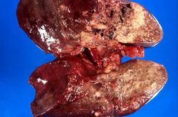 This gross photograph of the lungs shows areas of necrosis and consolidation.
 -
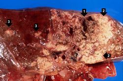 This higher-power view of the lung shows more clearly the areas of necrosis (1) and consolidation (2). Satellite lesions are also present (3).
 -
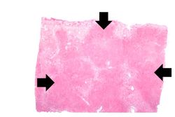 This is a low-power photomicrograph of lung showing many areas of consolidation (arrows).
 -
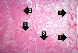 This is a high-power photomicrograph of lung section with pleura. The pleura (1) is thickened and contains inflammatory cells and fibrin. The areas of consolidation (2) are dense and filled with inflammatory cells.
 -
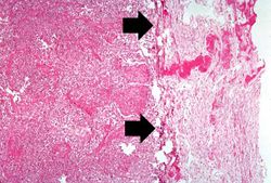 This is a higher-power photomicrograph of lung section with pleura. The pleura (arrows) is thickened and contains inflammatory cells and fibrin. The alveoli are filled with inflammatory cells. Some of the alveolar septa are congested with red blood cells.
 -
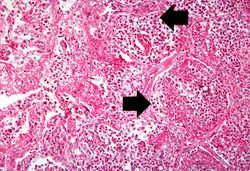 This high-power photomicrograph shows what appear to be inflammatory cells filling the alveoli. At this magnification, numerous round bodies (arrows) that look like inflammatory cell nuclei can be seen. However, on closer examination, some of these round bodies are surrounded by clear halos.
 -
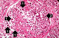 This high-power photomicrograph shows an alveolus filled with numerous round bodies up to 25 mm in diameter. Some of these double-contour bodies (1) have a dense center and a clear halo. These are the Blastomyces organisms. The typical B. dermatitides organism is smoothly-outlined with a central, densely basophilic cytoplasm surrounded by a clear halo. When stained with hematoxylin and eosin, the organism is outlined by a relatively thick cell wall. There are also numerous inflammatory cells (2) in the alveolus--neutrophils, lymphocytes and macrophages--which produce a pyogranulomatous inflammatory reaction.
 -
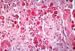 This is a high-power photomicrograph showing an alveolus filled with Blastomyces organisms. This section is stained with Periodic Acid-Schiff (PAS) to stain the Blastomyces organisms.
 -
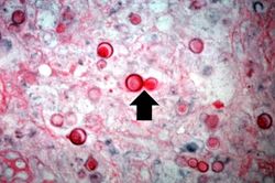 This high-power photomicrograph shows Blastomyces organisms stained with PAS. Note the budding organism (arrow). Blastomyces has a characteristic presentation of budding which aids in diagnosis of the fungus.
 -
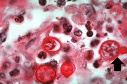 This is a very high-power photomicrograph showing Blastomyces organisms stained with PAS. Note the budding organism (arrow) and the underlying pyogranulomatous inflammatory reaction in the background.


Virtual Microscopy
H&E
PAS
Study Questions
Additional Resources
Reference
- eMedicine Medical Library: Imaging in Thoracic Blastomycosis
- eMedicine Medical Library: Blastomycosis
- Merck Manual: Blastomycosis
Journal Articles
- Rooney PJ, Klein BS. Linking fungal morphogenesis with virulence. Cell Microbiol 2002 Mar;4(3):127-37.
Images
| |||||