File:IPLab1FatNecrosis8.jpg: Difference between revisions
Jump to navigation
Jump to search
Seung Park (talk | contribs) No edit summary |
Peter Anderson uploaded a new version of File:IPLab1FatNecrosis8.jpg |
||
| (One intermediate revision by one other user not shown) | |||
| Line 1: | Line 1: | ||
This high-power photomicrograph demonstrates fat necrosis in the interlobular spaces of the pancreas. Note the granular blue-staining calcium deposits (arrows) within the fat cells. The clear areas represent artifact caused by the "washing-out" of fat from cells during tissue processing for histology. | |||


Latest revision as of 16:40, 27 June 2019
This high-power photomicrograph demonstrates fat necrosis in the interlobular spaces of the pancreas. Note the granular blue-staining calcium deposits (arrows) within the fat cells. The clear areas represent artifact caused by the "washing-out" of fat from cells during tissue processing for histology.
File history
Click on a date/time to view the file as it appeared at that time.
| Date/Time | Thumbnail | Dimensions | User | Comment | |
|---|---|---|---|---|---|
| current | 16:40, 27 June 2019 | 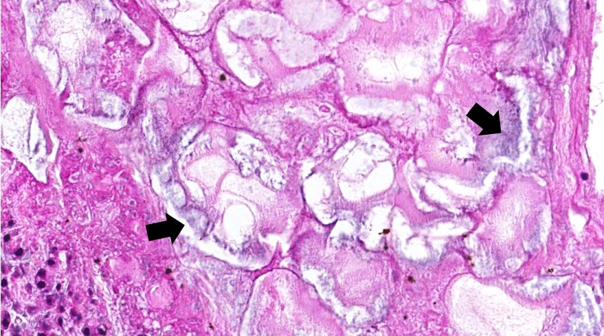 | 879 × 489 (166 KB) | Peter Anderson (talk | contribs) | |
| 20:18, 15 August 2013 | 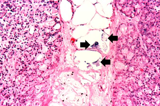 | 677 × 450 (72 KB) | Seung Park (talk | contribs) |
You cannot overwrite this file.
File usage
The following file is a duplicate of this file (more details):


There are no pages that use this file.
