Difference between revisions of "File:IPLab3Sarcoidosis3.jpg"
Seung Park (talk | contribs) (This is a photomicrograph of the small nodules (arrows) seen in the previous image. Close examination reveals that they are composed of large macrophages (epithelioid macrophages). These small granulomas form multiple series of reaction centers through...) |
(No difference)
|
Latest revision as of 03:30, 19 August 2013
This is a photomicrograph of the small nodules (arrows) seen in the previous image. Close examination reveals that they are composed of large macrophages (epithelioid macrophages). These small granulomas form multiple series of reaction centers throughout the lymph node. Note the remaining lymphocytes surrounding the granulomas.
File history
Click on a date/time to view the file as it appeared at that time.
| Date/Time | Thumbnail | Dimensions | User | Comment | |
|---|---|---|---|---|---|
| current | 03:30, 19 August 2013 | 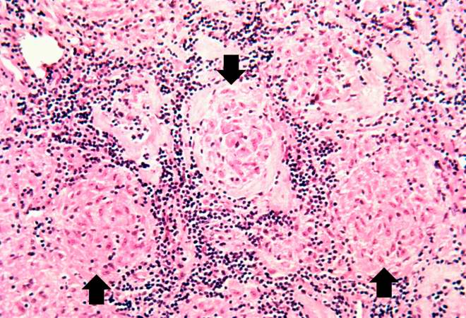 | 660 × 450 (84 KB) | Seung Park (talk | contribs) | This is a photomicrograph of the small nodules (arrows) seen in the previous image. Close examination reveals that they are composed of large macrophages (epithelioid macrophages). These small granulomas form multiple series of reaction centers through... |
- You cannot overwrite this file.
File usage
There are no pages that link to this file.