Difference between revisions of "File:IPLab3LobarPneumonia7.jpg"
Seung Park (talk | contribs) (This is another photomicrograph of the junction of the pleura (1) with the lung parenchyma. Note the alveoli on the left (2) filled with PMNs, alveolar macrophages, and fibrin. The dark red-stained material (3) in the center and left portions of the sl...) |
(No difference)
|
Latest revision as of 03:18, 19 August 2013
This is another photomicrograph of the junction of the pleura (1) with the lung parenchyma. Note the alveoli on the left (2) filled with PMNs, alveolar macrophages, and fibrin. The dark red-stained material (3) in the center and left portions of the slide is fibrin in the pleura. There are red blood cells trapped in the fibrinous pleuritis as well.
File history
Click on a date/time to view the file as it appeared at that time.
| Date/Time | Thumbnail | Dimensions | User | Comment | |
|---|---|---|---|---|---|
| current | 03:18, 19 August 2013 | 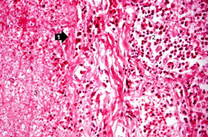 | 685 × 450 (78 KB) | Seung Park (talk | contribs) | This is another photomicrograph of the junction of the pleura (1) with the lung parenchyma. Note the alveoli on the left (2) filled with PMNs, alveolar macrophages, and fibrin. The dark red-stained material (3) in the center and left portions of the sl... |
- You cannot overwrite this file.
File usage
The following page links to this file: