Difference between revisions of "Cytologically Yours: Unknowns: 201312: Case 4"
(Created page with "==Clinical History== 25 year old female. Right breast core touch prep. ==Cytology== <gallery heights="250px" widths="250px"> CytologicallyYoursUnknowns201312-3-01.jpg Cytolog...") |
|||
| (One intermediate revision by the same user not shown) | |||
| Line 4: | Line 4: | ||
==Cytology== | ==Cytology== | ||
<gallery heights="250px" widths="250px"> | <gallery heights="250px" widths="250px"> | ||
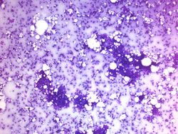
| − | CytologicallyYoursUnknowns201312- | + | CytologicallyYoursUnknowns201312-4-01.jpg |
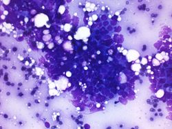
| − | CytologicallyYoursUnknowns201312- | + | CytologicallyYoursUnknowns201312-4-02.jpg |
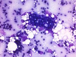
| − | CytologicallyYoursUnknowns201312- | + | CytologicallyYoursUnknowns201312-4-03.jpg |
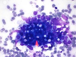
| − | CytologicallyYoursUnknowns201312- | + | CytologicallyYoursUnknowns201312-4-04.jpg |
| − | + | ||
| − | |||
</gallery> | </gallery> | ||
| Line 21: | Line 20: | ||
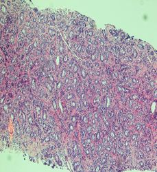
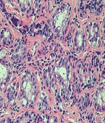
<gallery heights="250px" widths="250px"> | <gallery heights="250px" widths="250px"> | ||
| − | CytologicallyYoursUnknowns201312- | + | CytologicallyYoursUnknowns201312-4-05.jpg |
| − | CytologicallyYoursUnknowns201312- | + | CytologicallyYoursUnknowns201312-4-06.jpg |
</gallery> | </gallery> | ||
| + | |||
| + | |||
| + | ==Cytologic Features== | ||
| + | * The distinction between and adenoma and lactational change on cytology is extremely difficult (impossible). | ||
| + | * Hypercellar smears | ||
| + | * Loosely cohesive fragments | ||
| + | * Larger cells with prominent nucleoli | ||
| + | * Vacuolated cytoplasm | ||
| + | * Naked nuclei may be present in the background. | ||
| + | * Dirty background with inflammatory cells, macrophages, proteinaceous debris, blood | ||
| + | * May be initially confused with carcinoma. | ||
Latest revision as of 20:08, 10 January 2014
Contents
[hide]Clinical History
25 year old female. Right breast core touch prep.
Cytology
Resident Questions
Surgical Pathology
Cytologic Features
- The distinction between and adenoma and lactational change on cytology is extremely difficult (impossible).
- Hypercellar smears
- Loosely cohesive fragments
- Larger cells with prominent nucleoli
- Vacuolated cytoplasm
- Naked nuclei may be present in the background.
- Dirty background with inflammatory cells, macrophages, proteinaceous debris, blood
- May be initially confused with carcinoma.
