File:IPLab1Tuberculosis6.jpg: Difference between revisions
Jump to navigation
Jump to search
Seung Park (talk | contribs) No edit summary |
Seung Park (talk | contribs) No edit summary |
||
| Line 1: | Line 1: | ||
This photomicrograph shows a single nodule with an amorphous eosinophilic center and accumulations of cells around the outer edge. This is typical of a granuloma associated with tuberculosis in which there is a necrotic center (1) and a rim of lymphocytes, macrophages, and occasional multinucleated giant cells around the periphery. | |||


Latest revision as of 21:53, 15 August 2013
This photomicrograph shows a single nodule with an amorphous eosinophilic center and accumulations of cells around the outer edge. This is typical of a granuloma associated with tuberculosis in which there is a necrotic center (1) and a rim of lymphocytes, macrophages, and occasional multinucleated giant cells around the periphery.
File history
Click on a date/time to view the file as it appeared at that time.
| Date/Time | Thumbnail | Dimensions | User | Comment | |
|---|---|---|---|---|---|
| current | 21:50, 15 August 2013 | 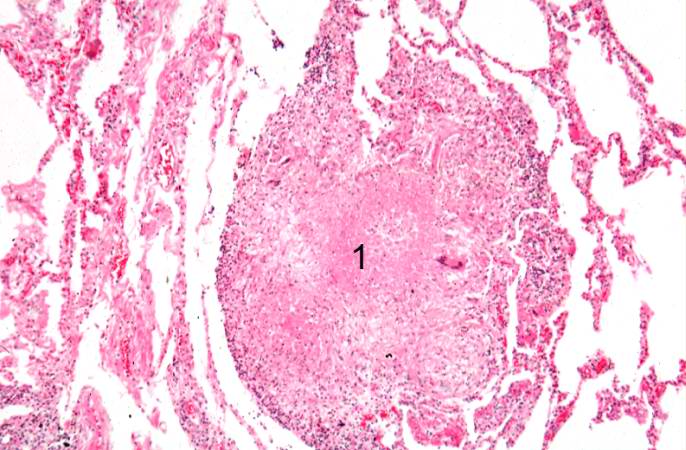 | 686 × 450 (64 KB) | Seung Park (talk | contribs) |
You cannot overwrite this file.
File usage
There are no pages that use this file.
