Difference between revisions of "File:IPLab1LungAbscess4.jpg"
Seung Park (talk | contribs) |
Seung Park (talk | contribs) |
||
| Line 1: | Line 1: | ||
| − | + | This higher-power photomicrograph of lung demonstrates the edge of the abscess. Note the loss of material from the center of the abscess (1) and loose necrotic material that has not been expelled (2). This material is made up of inflammatory cells (primarily dead white blood cells) and necrotic lung tissue. | |
Latest revision as of 16:20, 15 August 2013
This higher-power photomicrograph of lung demonstrates the edge of the abscess. Note the loss of material from the center of the abscess (1) and loose necrotic material that has not been expelled (2). This material is made up of inflammatory cells (primarily dead white blood cells) and necrotic lung tissue.
An abscess is a collection of pus (white blood cells) within a cavity formed by disintegrated tissue.
File history
Click on a date/time to view the file as it appeared at that time.
| Date/Time | Thumbnail | Dimensions | User | Comment | |
|---|---|---|---|---|---|
| current | 16:15, 15 August 2013 | 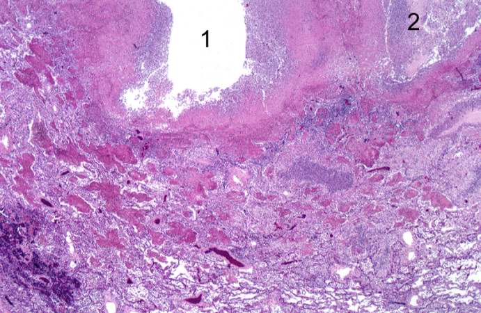 | 691 × 450 (75 KB) | Seung Park (talk | contribs) |
- You cannot overwrite this file.
File usage
There are no pages that link to this file.