Difference between revisions of "File:IPLab13Hyaline6.jpg"
Seung Park (talk | contribs) (This is a medium-power photomicrograph showing a large bronchus with cartilage. Interstitial congestion with numerous red cells is apparent. Even at this magnification hyaline membranes (arrows) can be seen lining the alveoli.) |
(No difference)
|
Latest revision as of 05:51, 21 August 2013
This is a medium-power photomicrograph showing a large bronchus with cartilage. Interstitial congestion with numerous red cells is apparent. Even at this magnification hyaline membranes (arrows) can be seen lining the alveoli.
File history
Click on a date/time to view the file as it appeared at that time.
| Date/Time | Thumbnail | Dimensions | User | Comment | |
|---|---|---|---|---|---|
| current | 05:51, 21 August 2013 | 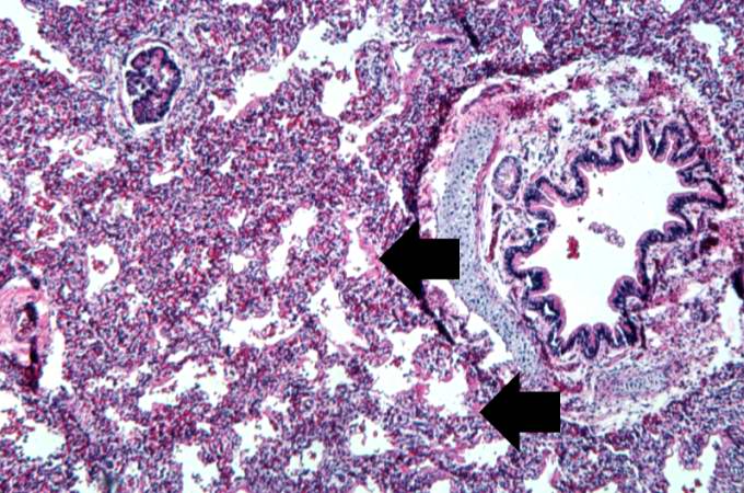 | 680 × 450 (85 KB) | Seung Park (talk | contribs) | This is a medium-power photomicrograph showing a large bronchus with cartilage. Interstitial congestion with numerous red cells is apparent. Even at this magnification hyaline membranes (arrows) can be seen lining the alveoli. |
- You cannot overwrite this file.
File usage
The following page links to this file: