Difference between revisions of "File:IPLab12Mesothelioma5.jpg"
Seung Park (talk | contribs) (This higher-power photomicrograph of left lung shows areas of consolidation and fibrosis (arrows). Also note that in many of these areas there are clusters of blue cells.) |
(No difference)
|
Latest revision as of 05:33, 21 August 2013
This higher-power photomicrograph of left lung shows areas of consolidation and fibrosis (arrows). Also note that in many of these areas there are clusters of blue cells.
File history
Click on a date/time to view the file as it appeared at that time.
| Date/Time | Thumbnail | Dimensions | User | Comment | |
|---|---|---|---|---|---|
| current | 05:33, 21 August 2013 | 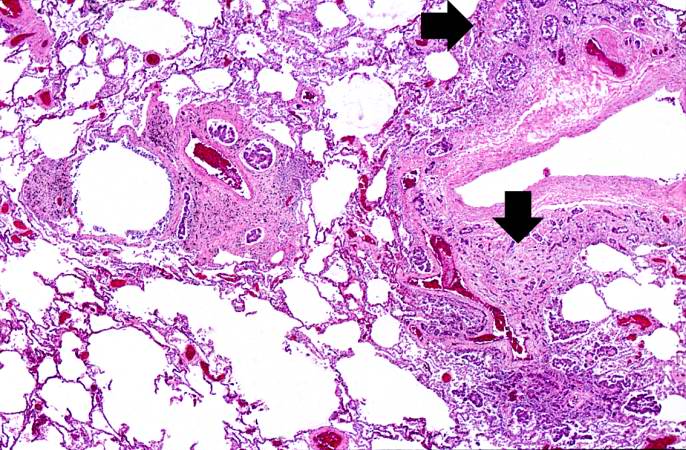 | 686 × 450 (99 KB) | Seung Park (talk | contribs) | This higher-power photomicrograph of left lung shows areas of consolidation and fibrosis (arrows). Also note that in many of these areas there are clusters of blue cells. |
- You cannot overwrite this file.
File usage
The following page links to this file: