Difference between revisions of "File:IPLab12Mesothelioma1.jpg"
Seung Park (talk | contribs) (This is a gross photograph of the lungs removed at autopsy. There is thickening of the pleural surface due to the tumor (arrows). The apical portion of the left lobe of the lung was ripped while trying to sever the adhesions between the lung and the ch...) |
(No difference)
|
Latest revision as of 05:32, 21 August 2013
This is a gross photograph of the lungs removed at autopsy. There is thickening of the pleural surface due to the tumor (arrows). The apical portion of the left lobe of the lung was ripped while trying to sever the adhesions between the lung and the chest wall.
File history
Click on a date/time to view the file as it appeared at that time.
| Date/Time | Thumbnail | Dimensions | User | Comment | |
|---|---|---|---|---|---|
| current | 05:32, 21 August 2013 | 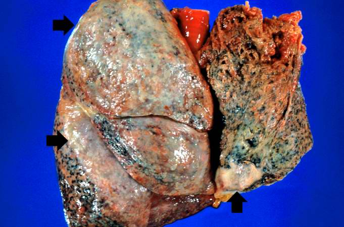 | 681 × 450 (52 KB) | Seung Park (talk | contribs) | This is a gross photograph of the lungs removed at autopsy. There is thickening of the pleural surface due to the tumor (arrows). The apical portion of the left lobe of the lung was ripped while trying to sever the adhesions between the lung and the ch... |
- You cannot overwrite this file.
File usage
The following page links to this file: