Difference between revisions of "File:IPLab12RadiationFibrosis10.jpg"
Seung Park (talk | contribs) (This is another high-power photomicrograph of an area of tissue with diffuse fibrosis and thickening of the alveolar septa. There are also accumulations of anthracotic pigment in this area (arrows).) |
(No difference)
|
Latest revision as of 05:25, 21 August 2013
This is another high-power photomicrograph of an area of tissue with diffuse fibrosis and thickening of the alveolar septa. There are also accumulations of anthracotic pigment in this area (arrows).
Anthracotic pigment is coal dust deposited in the lungs--it is seen in coal miners, city-dwellers, and smokers.
File history
Click on a date/time to view the file as it appeared at that time.
| Date/Time | Thumbnail | Dimensions | User | Comment | |
|---|---|---|---|---|---|
| current | 05:25, 21 August 2013 | 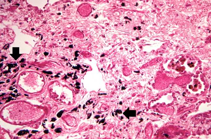 | 681 × 450 (80 KB) | Seung Park (talk | contribs) | This is another high-power photomicrograph of an area of tissue with diffuse fibrosis and thickening of the alveolar septa. There are also accumulations of anthracotic pigment in this area (arrows). |
- You cannot overwrite this file.
File usage
The following page links to this file: