Difference between revisions of "File:IPLab11Schistosomiasis3.jpg"
Seung Park (talk | contribs) (This is a photomicrograph of the liver from another patient with Schistosoma mansoni. These eggs have elicited a robust inflammatory response, including a multinucleated giant cell. These lesions go on to heal by fibrosis resulting in cirrhosis and the...) |
(No difference)
|
Latest revision as of 05:05, 21 August 2013
This is a photomicrograph of the liver from another patient with Schistosoma mansoni. These eggs have elicited a robust inflammatory response, including a multinucleated giant cell. These lesions go on to heal by fibrosis resulting in cirrhosis and the classic "pipe-stem" fibrosis.
Cirrhosis is a liver disease characterized by necrosis, fibrosis, loss of normal liver architecture, and hyperplastic nodules.
File history
Click on a date/time to view the file as it appeared at that time.
| Date/Time | Thumbnail | Dimensions | User | Comment | |
|---|---|---|---|---|---|
| current | 05:05, 21 August 2013 | 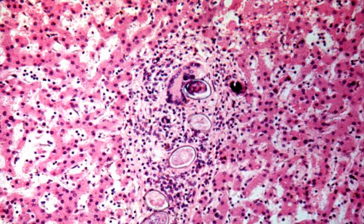 | 729 × 450 (79 KB) | Seung Park (talk | contribs) | This is a photomicrograph of the liver from another patient with Schistosoma mansoni. These eggs have elicited a robust inflammatory response, including a multinucleated giant cell. These lesions go on to heal by fibrosis resulting in cirrhosis and the... |
- You cannot overwrite this file.
File usage
The following page links to this file: