Difference between revisions of "File:IPLab11Leishmaniasis6.jpg"
Seung Park (talk | contribs) (This is a high-power photomicrograph of a touch prep made from the skin lesion at the time of biopsy. A single macrophage can be seen with intracytoplasmic leishmania organisms (arrows).) |
(No difference)
|
Latest revision as of 04:58, 21 August 2013
This is a high-power photomicrograph of a touch prep made from the skin lesion at the time of biopsy. A single macrophage can be seen with intracytoplasmic leishmania organisms (arrows).
File history
Click on a date/time to view the file as it appeared at that time.
| Date/Time | Thumbnail | Dimensions | User | Comment | |
|---|---|---|---|---|---|
| current | 04:58, 21 August 2013 | 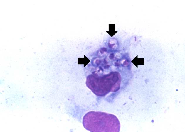 | 630 × 450 (19 KB) | Seung Park (talk | contribs) | This is a high-power photomicrograph of a touch prep made from the skin lesion at the time of biopsy. A single macrophage can be seen with intracytoplasmic leishmania organisms (arrows). |
- You cannot overwrite this file.
File usage
The following page links to this file: