Difference between revisions of "File:IPLab10Crypto2.jpg"
Seung Park (talk | contribs) (This is a gross photomicrograph of this lung taken at autopsy. Note the areas of emphysema (1) and consolidation (2).) |
(No difference)
|
Latest revision as of 04:10, 21 August 2013
This is a gross photomicrograph of this lung taken at autopsy. Note the areas of emphysema (1) and consolidation (2).
Pulmonary emphysema is a condition in which the air spaces distal to the terminal bronchioles are permanently increased in size due to either destruction of the wall or alveolar dilatation.
File history
Click on a date/time to view the file as it appeared at that time.
| Date/Time | Thumbnail | Dimensions | User | Comment | |
|---|---|---|---|---|---|
| current | 04:10, 21 August 2013 | 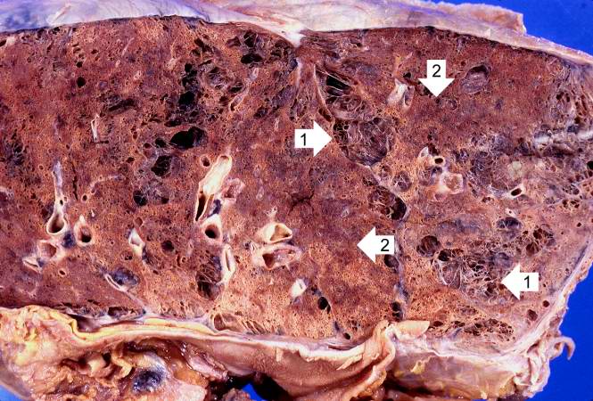 | 665 × 450 (83 KB) | Seung Park (talk | contribs) | This is a gross photomicrograph of this lung taken at autopsy. Note the areas of emphysema (1) and consolidation (2). |
- You cannot overwrite this file.
File usage
The following page links to this file: