Difference between revisions of "File:IPLab10Candidiasis7.jpg"
Seung Park (talk | contribs) (This is a low-power photomicrograph of the kidney from this same case. Note the Candida colonies (arrows). The pseudohyphae are evident around the periphery of these colonies even at this low magnification.) |
(No difference)
|
Latest revision as of 04:03, 21 August 2013
This is a low-power photomicrograph of the kidney from this same case. Note the Candida colonies (arrows). The pseudohyphae are evident around the periphery of these colonies even at this low magnification.
File history
Click on a date/time to view the file as it appeared at that time.
| Date/Time | Thumbnail | Dimensions | User | Comment | |
|---|---|---|---|---|---|
| current | 04:03, 21 August 2013 | 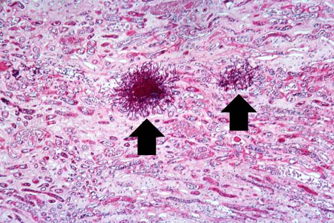 | 673 × 450 (77 KB) | Seung Park (talk | contribs) | This is a low-power photomicrograph of the kidney from this same case. Note the Candida colonies (arrows). The pseudohyphae are evident around the periphery of these colonies even at this low magnification. |
- You cannot overwrite this file.
File usage
The following page links to this file: