File:IPLab5Downs1.jpg
No higher resolution available.
IPLab5Downs1.jpg (596 × 407 pixels, file size: 18 KB, MIME type: image/jpeg)
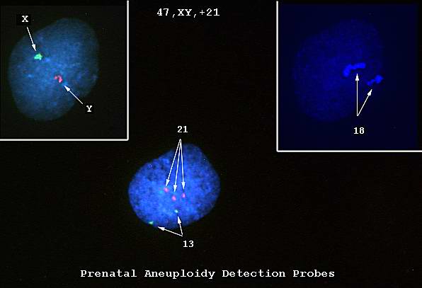
This is a photomicrograph of cells obtained by amniocentesis that were stained using FISH. The cell in panel 1 was stained with markers specific for the X and Y-chromosomes. The cell in panel 2 was stained with a marker specific for chromosome 18. The cell in the center was stained with markers for chromosomes 13 and 21. Note that there are three copies of chromosome 21.
File history
Click on a date/time to view the file as it appeared at that time.
| Date/Time | Thumbnail | Dimensions | User | Comment | |
|---|---|---|---|---|---|
| current | 13:27, 19 August 2013 | | 596 × 407 (18 KB) | Seung Park (talk | contribs) | This is a photomicrograph of cells obtained by amniocentesis that were stained using FISH. The cell in panel 1 was stained with markers specific for the X and Y-chromosomes. The cell in panel 2 was stained with a marker specific for chromosome 18. The ... |
You cannot overwrite this file.
File usage
The following page uses this file:
