File:IPLab3Tuberculosis1.jpg
No higher resolution available.
IPLab3Tuberculosis1.jpg (698 × 450 pixels, file size: 51 KB, MIME type: image/jpeg)
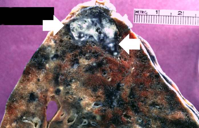
This is a gross photograph of a lung containing a nodular lesion at the lung apex (arrows). Note that the lesion appears solid and has a whitish coloration indicating considerable fibrous connective tissue. This is a healed granuloma due to primary tuberculosis in the lung. There are smaller focal lesions adjacent to the major mass. In addition, note the extensive anthracosis in this lung.
File history
Click on a date/time to view the file as it appeared at that time.
| Date/Time | Thumbnail | Dimensions | User | Comment | |
|---|---|---|---|---|---|
| current | 22:38, 18 August 2013 | | 698 × 450 (51 KB) | Seung Park (talk | contribs) | This is a gross photograph of a lung containing a nodular lesion at the lung apex (arrows). Note that the lesion appears solid and has a whitish coloration indicating considerable fibrous connective tissue. This is a healed granuloma due to primary tub... |
You cannot overwrite this file.
File usage
The following page uses this file:
