IPLab:Lab 3:Sarcoidosis
Clinical Summary
This 33-year-old white female was admitted for evaluation of abnormal findings on a chest x-ray. She was asymptomatic and a physical examination revealed no significant abnormalities. Laboratory results indicated hypercalcemia and elevated gamma globulin. Radiographic examination showed enlarged subcarinal, hilar, and right paratracheal lymph nodes. A right paratracheal lymph node was biopsied. Special stains for acid-fast bacilli and fungi were negative and a diagnosis of sarcoidosis was made.
Images
-
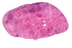 This is a low-power photomicrograph of a lymph node. Note the rather pale-pink color of the tissue with dark-staining cells found in only a few scattered areas. These darker cells represent the original lymphocytes of this lymphoid organ.
 -
 This photomicrograph of lymph node tissue illustrates a paucity of lymphocytes as well as numerous small, pale-staining nodules (arrows) throughout the tissue.
 -
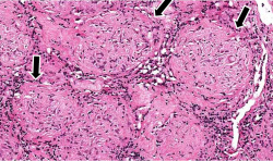 This is a photomicrograph of the small nodules (arrows) seen in the previous image. Close examination reveals that they are composed of large macrophages (epithelioid macrophages). These small granulomas form multiple series of reaction centers throughout the lymph node. Note the remaining lymphocytes surrounding the granulomas.
 -
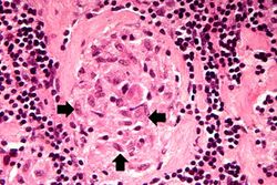 This photomicrograph of a single granuloma illustrates the individual macrophages (arrows) which make up the bulk of this tissue. There is an absence of necrosis in the center of the lesions in this case.
 -
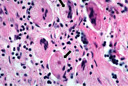 This is a photomicrograph of a multinucleated giant cell (1). In the center of this foreign body-containing giant cell there is a small asteroid body (2). There is no functional significance to this asteroid body.


Virtual Microscopy
Lymph Node: Sarcoidosis
Lymph Node
Study Questions
Additional Resources
Reference
- eMedicine Medical Library: Acute Complications of Sarcoidosis
- eMedicine Medical Library: Sarcoidosis
- eMedicine Medical Library: Dermatologic Manifestations of Sarcoidosis
- eMedicine Medical Library: Neurosarcoidosis
- Merck Manual: Sarcoidosis
Journal Articles
- Drent M, Crouser ED, Grunewald J. Challenges of Sarcoidosis and Its Management. N Engl J Med 2021; 385:1018-1032
- English JC, Patel PJ, Greer KE. Sarcoidosis. J Am Acad Dermatol 2001;44:725-43
- Johns CJ, Michele TM. The clinical management of sarcoidosis. A 50-year experience at the Johns Hopkins Hospital. Medicine (Baltimore) 1999 Mar;78(2):65-111.