Cytologically Yours: CoW: 20140113
Jump to navigation
Jump to search
Clinical Summary
The patient is a 69 year old female with a new mass in the right pulmonary hilum.
Past Medical History
- Hyperlipidemia
- Osteoporosis
- Chronic obstructive pulmonary disease
- Hepatitis
- Smoker (1 pack per day for 40 years, quit in 2002)
Past Surgical History
- Hysterectomy (1975)
- Hernia repair (1992)
- Cardiac catheterization (2002)
- Lumbarsacral discectomy (2008)
- Lumbarsacral fusion with rod placement (2013)
CT
- 4.8 x 3.1 cm mass in the right hilum.
- 4.4 x 1.5 cm nodular densityin the right upper lobe.
- 1.5 cm enlarged paratracheal lymph node.
- CT with contrast: mass in the right hilum extends into the mediastium and the SVC is compressed.
Clinical Plan
Bronchoscopy for furhter evaluation.
Pathology
Cytology
-
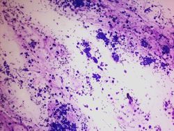 10x magnification of a cellular specimen.(DQ)
 -
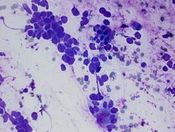 40x magnification showing cohesive groups of atypical cells adjacent to bronchial cells. (DQ)
 -
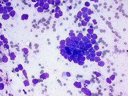 40x magnification showing a large group of cohesive cells that are molding upon one another. (DQ)
 -
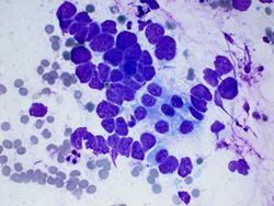 40x magnification showing cells that are molding and forming a row of cells.(DQ)
 -
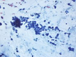 20x magnification of cells with chromatin that looks punctate.(pap)
 -
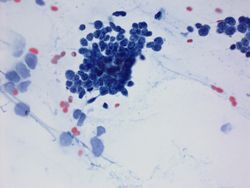 60x magnification of cells with punctate chromatin and molding.(pap)


Resident Questions
Final Diagnosis
Cytology
- Small Cell Carcinoma.
Discussion
Small cell carcinoma is associated with smoking and is generally a central lesion that arises in larger airways. Small cell carcinoma is prone to central necrosis and radiologically this may be seen.
| ||||||||