File:IPLab3HealedMyocardialInfarction6.jpg
No higher resolution available.
IPLab3HealedMyocardialInfarction6.jpg (668 × 450 pixels, file size: 70 KB, MIME type: image/jpeg)
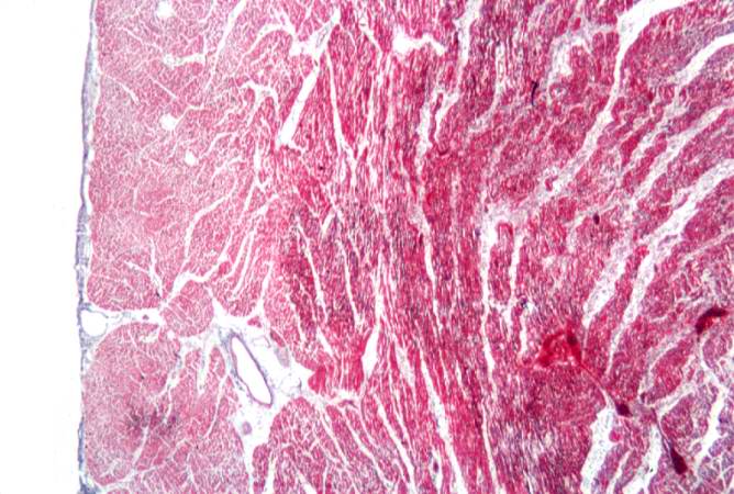
This is a photomicrograph of a trichrome-stained section from a heart with an acute myocardial infarction. Note that there is little fibrous connective tissue. It is too early for scar formation to have taken place in this acute lesion.
File history
Click on a date/time to view the file as it appeared at that time.
| Date/Time | Thumbnail | Dimensions | User | Comment | |
|---|---|---|---|---|---|
| current | 23:38, 18 August 2013 | | 668 × 450 (70 KB) | Seung Park (talk | contribs) | This is a photomicrograph of a trichrome-stained section from a heart with an acute myocardial infarction. Note that there is little fibrous connective tissue. It is too early for scar formation to have taken place in this acute lesion. |
You cannot overwrite this file.
File usage
There are no pages that use this file.
