File:IPLab3LobarPneumonia4.jpg
IPLab3LobarPneumonia4.jpg (731 × 450 pixels, file size: 31 KB, MIME type: image/jpeg)
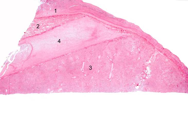
This low-power photomicrograph of the lung shows: (1) markedly thickened pleura, indicating an inflammatory process which has been present for several days; (2) a small wedge-shaped segment of normal lung which is somewhat compressed due to artifact; (3) a large wedge-shaped segment of lung tissue with lobar pneumonia, which appears very dense due to extensive cellular infiltrate; and (4) a greatly thickened pleural space between these two lung lobes. This interlobar space is markedly thickened due to deposition of fibrinous exudate.
File history
Click on a date/time to view the file as it appeared at that time.
| Date/Time | Thumbnail | Dimensions | User | Comment | |
|---|---|---|---|---|---|
| current | 22:17, 18 August 2013 | | 731 × 450 (31 KB) | Seung Park (talk | contribs) | This low-power photomicrograph of the lung shows: (1) markedly thickened pleura, indicating an inflammatory process which has been present for several days; (2) a small wedge-shaped segment of normal lung which is somewhat compressed due to artifact; (... |
You cannot overwrite this file.
File usage
The following page uses this file:
