-
This is a gross photograph of thyroid gland taken at autopsy. The gland is only slightly enlarged and has a firm texture.
-
This is a low-power photomicrograph of thyroid from this case. Note that the tissue is more cellular than one would expect and there does not appear to be normal colloid-filled blue spaces in this gland.
-
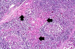
This is a higher-power photomicrograph of thyroid from this case. Note the large number of blue-staining inflammatory cells in this tissue. These cells appear to be forming germinal centers. Some residual thyroid gland tissue can be seen in this section (arrows).
-
This is another view of thyroid gland filled with inflammatory cells forming germinal centers (arrows).
-
This is a higher-power photomicrograph of thyroid from this case showing the inflammatory cells and the residual thyroid tissue.
-
This is another higher-power photomicrograph of thyroid from this case showing the inflammatory cells and the residual thyroid tissue.
-
This is a high-power photomicrograph showing the inflammatory cells infiltrating into the residual thyroid tissue (arrows).
-
This is a high-power photomicrograph showing the lymphocytes and plasma cells surrounding the thyroid gland epithelium.
-
This high-power photomicrograph shows more clearly the lymphocytes and plasma cells surrounding the thyroid gland epithelium. Large, eosinophilic, degenerating thyroid gland cells (Hurthle cells) can be seen in this section (arrows).
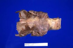 This is a gross photograph of thyroid gland taken at autopsy. The gland is only slightly enlarged and has a firm texture.
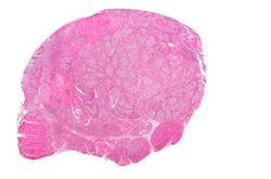 This is a low-power photomicrograph of thyroid from this case. Note that the tissue is more cellular than one would expect and there does not appear to be normal colloid-filled blue spaces in this gland.
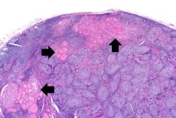 This is a higher-power photomicrograph of thyroid from this case. Note the large number of blue-staining inflammatory cells in this tissue. These cells appear to be forming germinal centers. Some residual thyroid gland tissue can be seen in this section (arrows).
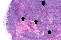 This is another view of thyroid gland filled with inflammatory cells forming germinal centers (arrows).
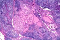 This is a higher-power photomicrograph of thyroid from this case showing the inflammatory cells and the residual thyroid tissue.
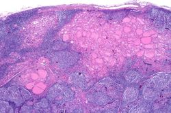 This is another higher-power photomicrograph of thyroid from this case showing the inflammatory cells and the residual thyroid tissue.
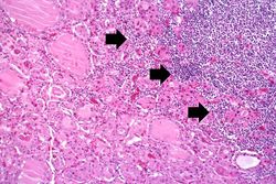 This is a high-power photomicrograph showing the inflammatory cells infiltrating into the residual thyroid tissue (arrows).
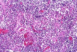 This is a high-power photomicrograph showing the lymphocytes and plasma cells surrounding the thyroid gland epithelium.
 This high-power photomicrograph shows more clearly the lymphocytes and plasma cells surrounding the thyroid gland epithelium. Large, eosinophilic, degenerating thyroid gland cells (Hurthle cells) can be seen in this section (arrows).


