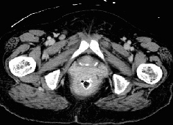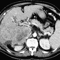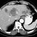
RADIOLOGY: GASTROINTESTINAL: GI: Case# 32867: SQAMOUS CELL ANAL/RECTAL CA. 69 year old black female with rectal mass. There is circumferential rectal wall thickening extending from the region of the anus to the rectosigmoid junction. There are extensive strandy changes surrounding the rectum and extending anteriorly to abut the uterus and vagina. No definite pelvic lymphadenopathy is seen. Colorectal cancer is the third most common cause of cancer death in the United States. It appears to be associated with diets high in animal fat and protein and low in fiber. Seventy percent of colorectal cancers occur in the rectosigmoid region. Rectal cancers may present with bleeding, change in bowel habits, perineal pain, and symptoms of invasion of adjacent organs including hematuria, urinary frequency, or vaginal fistulas. Surgical removal is the only effective therapy. Anterior resection with anastomosis to the rectal stump is performed for upper rectal tumors and a combined abdominal and perineal resection with a permanent colostomy for distal rectal lesions. On CT, rectal lesions may appear as focal lobulated soft tissue masses with asymmetrical or circumferential thickening of the bowel wall. Narrowing of the lumen and bowel obstruction may be evident. Extension into pericolonic fat or adjacent structures, regional adenopathy, adrenal or liver metastases, and hydronephrosis may also be visualized. Common sites of local extension include the pelvic musculature, bladder, prostate, ovaries, and seminal vesicles. Although CT is not as accurate in staging colorectal cancer, predicting local extension, or detecting spread to lymph nodes, it is the procedure of choice for detecting recurrence of carcinoma, particularly in patients who have undergone abdominoperineal resection.
- Author
- Peter Anderson
- Posted on
- Thursday 1 August 2013
- Tags
- gastrointestinal, radiology
- Albums
- Visits
- 1074


0 comments