File:IPLab6PAN12.jpg
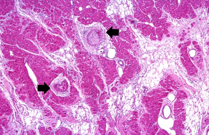
Revision as of 18:00, 20 August 2013 by Peter Anderson (talk | contribs) (This is a higher-power photomicrograph of the affected vessels in the heart (arrows). There are areas of fibrosis (old infarcts) in the myocardium adjacent to these affected vessels.)
No higher resolution available.
IPLab6PAN12.jpg (697 × 450 pixels, file size: 88 KB, MIME type: image/jpeg)
This is a higher-power photomicrograph of the affected vessels in the heart (arrows). There are areas of fibrosis (old infarcts) in the myocardium adjacent to these affected vessels.
File history
Click on a date/time to view the file as it appeared at that time.
| Date/Time | Thumbnail | Dimensions | User | Comment | |
|---|---|---|---|---|---|
| current | 18:00, 20 August 2013 | | 697 × 450 (88 KB) | Peter Anderson (talk | contribs) | This is a higher-power photomicrograph of the affected vessels in the heart (arrows). There are areas of fibrosis (old infarcts) in the myocardium adjacent to these affected vessels. |
- You cannot overwrite this file.
File usage
The following page links to this file: