File:IPLab6PAN11.jpg
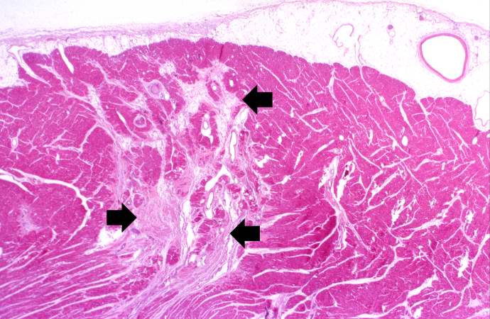
Revision as of 17:59, 20 August 2013 by Peter Anderson (talk | contribs) (This is a low-power photomicrograph of the heart. There are areas of fibrosis in the myocardium (arrows). Note that the large epicardial coronary artery is normal.)
No higher resolution available.
IPLab6PAN11.jpg (690 × 450 pixels, file size: 69 KB, MIME type: image/jpeg)
This is a low-power photomicrograph of the heart. There are areas of fibrosis in the myocardium (arrows). Note that the large epicardial coronary artery is normal.
File history
Click on a date/time to view the file as it appeared at that time.
| Date/Time | Thumbnail | Dimensions | User | Comment | |
|---|---|---|---|---|---|
| current | 17:59, 20 August 2013 | | 690 × 450 (69 KB) | Peter Anderson (talk | contribs) | This is a low-power photomicrograph of the heart. There are areas of fibrosis in the myocardium (arrows). Note that the large epicardial coronary artery is normal. |
- You cannot overwrite this file.
File usage
The following page links to this file: