File:IPLab5Hemochromatosis4.jpg
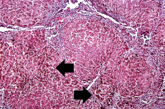
Revision as of 14:53, 20 August 2013 by Peter Anderson (talk | contribs) (This higher-power view of liver from this case demonstrates the nodules and the brown/black pigment within liver parenchymal cells (arrows).)
No higher resolution available.
IPLab5Hemochromatosis4.jpg (679 × 450 pixels, file size: 94 KB, MIME type: image/jpeg)
This higher-power view of liver from this case demonstrates the nodules and the brown/black pigment within liver parenchymal cells (arrows).
File history
Click on a date/time to view the file as it appeared at that time.
| Date/Time | Thumbnail | Dimensions | User | Comment | |
|---|---|---|---|---|---|
| current | 14:53, 20 August 2013 | | 679 × 450 (94 KB) | Peter Anderson (talk | contribs) | This higher-power view of liver from this case demonstrates the nodules and the brown/black pigment within liver parenchymal cells (arrows). |
- You cannot overwrite this file.
File usage
The following page links to this file: