File:IPLab2Calcification4.jpg
Revision as of 16:34, 19 August 2013 by Peter Anderson (talk | contribs) (This high-power photomicrograph of a blood vessel shows calcium deposits in the vascular wall (1) and proteinaceous material (2) (from edema) within some of the alveoli. The smooth muscle in the vessel wall has been almost completely replaced by calciu...)
No higher resolution available.
IPLab2Calcification4.jpg (679 × 450 pixels, file size: 65 KB, MIME type: image/jpeg)
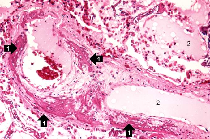
This high-power photomicrograph of a blood vessel shows calcium deposits in the vascular wall (1) and proteinaceous material (2) (from edema) within some of the alveoli. The smooth muscle in the vessel wall has been almost completely replaced by calcium deposits.
File history
Click on a date/time to view the file as it appeared at that time.
| Date/Time | Thumbnail | Dimensions | User | Comment | |
|---|---|---|---|---|---|
| current | 16:34, 19 August 2013 | | 679 × 450 (65 KB) | Peter Anderson (talk | contribs) | This high-power photomicrograph of a blood vessel shows calcium deposits in the vascular wall (1) and proteinaceous material (2) (from edema) within some of the alveoli. The smooth muscle in the vessel wall has been almost completely replaced by calciu... |
- You cannot overwrite this file.
File usage
The following page links to this file: