File:IPLab2Calcification3.jpg
Revision as of 16:33, 19 August 2013 by Peter Anderson (talk | contribs) (A higher-power photomicrograph shows a blood vessel cut in longitudinal section (1). Several of the alveoli are filled with a pink-staining proteinaceous fluid (2) indicative of pulmonary edema. The alveolar septa and the wall of the blood vessel have ...)
No higher resolution available.
IPLab2Calcification3.jpg (677 × 450 pixels, file size: 78 KB, MIME type: image/jpeg)
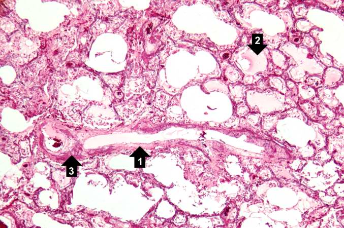
A higher-power photomicrograph shows a blood vessel cut in longitudinal section (1). Several of the alveoli are filled with a pink-staining proteinaceous fluid (2) indicative of pulmonary edema. The alveolar septa and the wall of the blood vessel have a purplish color due to massive deposition of mineral (primarily calcium) in these tissues (3).
Pulmonary edema refers to the accumulation of fluid in the pulmonary alveolar and tissue spaces as a result of changes in capillary permeability and/or increases in capillary hydrostatic pressure.
File history
Click on a date/time to view the file as it appeared at that time.
| Date/Time | Thumbnail | Dimensions | User | Comment | |
|---|---|---|---|---|---|
| current | 16:33, 19 August 2013 | | 677 × 450 (78 KB) | Peter Anderson (talk | contribs) | A higher-power photomicrograph shows a blood vessel cut in longitudinal section (1). Several of the alveoli are filled with a pink-staining proteinaceous fluid (2) indicative of pulmonary edema. The alveolar septa and the wall of the blood vessel have ... |
- You cannot overwrite this file.
File usage
The following page links to this file: