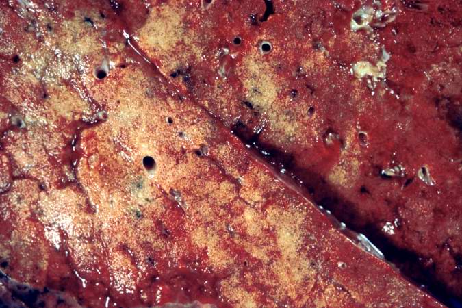
File:IPLab2Calcification1.jpg
Revision as of 16:32, 19 August 2013 by Peter Anderson (talk | contribs) (This is a gross photograph of the cut section of the patient's lung showing evidence of severe metastatic calcification. The lung tissue has a rough, firm appearance with open airways.)
No higher resolution available.
IPLab2Calcification1.jpg (675 × 450 pixels, file size: 72 KB, MIME type: image/jpeg)
This is a gross photograph of the cut section of the patient's lung showing evidence of severe metastatic calcification. The lung tissue has a rough, firm appearance with open airways.
The deposition of calcium in normal tissues as a result of elevations in blood calcium.
File history
Click on a date/time to view the file as it appeared at that time.
| Date/Time | Thumbnail | Dimensions | User | Comment | |
|---|---|---|---|---|---|
| current | 16:32, 19 August 2013 | | 675 × 450 (72 KB) | Peter Anderson (talk | contribs) | This is a gross photograph of the cut section of the patient's lung showing evidence of severe metastatic calcification. The lung tissue has a rough, firm appearance with open airways. |
- You cannot overwrite this file.
File usage
The following page links to this file: