No higher resolution available.
IPLab1FatNecrosis1.jpg (681 × 450 pixels, file size: 36 KB, MIME type: image/jpeg)
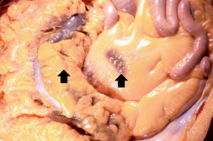
This gross photograph shows the intestines and omentum at autopsy. Note the small (5-15 mm in diameter) white nodules on the surface of the omental and mesenteric fat tissue (arrows).
File history
Click on a date/time to view the file as it appeared at that time.
| Date/Time | Thumbnail | Dimensions | User | Comment | |
|---|---|---|---|---|---|
| current | 01:17, 16 August 2013 | | 681 × 450 (36 KB) | Seung Park (talk | contribs) |
- You cannot overwrite this file.
File usage
The following page links to this file: