File:IPLab13WT1.jpg
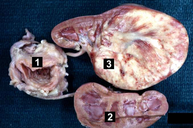
Revision as of 05:54, 21 August 2013 by Seung Park (talk | contribs) (This is a gross photograph of a bladder (1) to which are attached a normal kidney (2) and a kidney with Wilms' tumor (3). A large mass extends from the superior pole of the affected kidney. The renal capsule can be seen extending around this tumor.)
No higher resolution available.
IPLab13WT1.jpg (678 × 450 pixels, file size: 51 KB, MIME type: image/jpeg)
This is a gross photograph of a bladder (1) to which are attached a normal kidney (2) and a kidney with Wilms' tumor (3). A large mass extends from the superior pole of the affected kidney. The renal capsule can be seen extending around this tumor.
File history
Click on a date/time to view the file as it appeared at that time.
| Date/Time | Thumbnail | Dimensions | User | Comment | |
|---|---|---|---|---|---|
| current | 05:54, 21 August 2013 | | 678 × 450 (51 KB) | Seung Park (talk | contribs) | This is a gross photograph of a bladder (1) to which are attached a normal kidney (2) and a kidney with Wilms' tumor (3). A large mass extends from the superior pole of the affected kidney. The renal capsule can be seen extending around this tumor. |
- You cannot overwrite this file.
File usage
The following page links to this file: