File:IPLab12Acetaminophen4.jpg
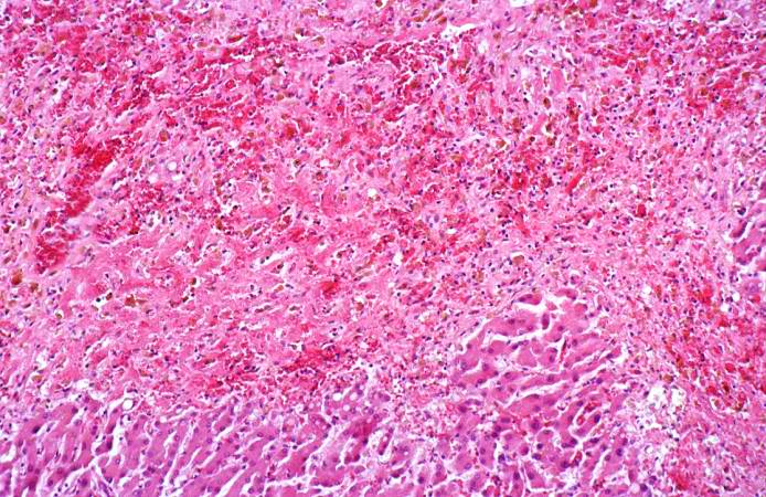
Revision as of 05:19, 21 August 2013 by Seung Park (talk | contribs) (This is a medium-power photomicrograph of the areas of hemorrhagic necrosis. Note the coagulation necrosis and hemorrhage in this area. Viable hepatocytes can be seen along the edge of this lesion.)
No higher resolution available.
IPLab12Acetaminophen4.jpg (694 × 450 pixels, file size: 95 KB, MIME type: image/jpeg)
This is a medium-power photomicrograph of the areas of hemorrhagic necrosis. Note the coagulation necrosis and hemorrhage in this area. Viable hepatocytes can be seen along the edge of this lesion.
File history
Click on a date/time to view the file as it appeared at that time.
| Date/Time | Thumbnail | Dimensions | User | Comment | |
|---|---|---|---|---|---|
| current | 05:19, 21 August 2013 | | 694 × 450 (95 KB) | Seung Park (talk | contribs) | This is a medium-power photomicrograph of the areas of hemorrhagic necrosis. Note the coagulation necrosis and hemorrhage in this area. Viable hepatocytes can be seen along the edge of this lesion. |
- You cannot overwrite this file.
File usage
The following page links to this file: