File:IPLab6PAN7.jpg
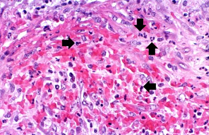
Revision as of 17:57, 20 August 2013 by Peter Anderson (talk | contribs) (This is a high-power photomicrograph of the vessel wall. There is hemorrhage and infiltration with inflammatory cells--primarily neutrophils (arrows).)
No higher resolution available.
IPLab6PAN7.jpg (697 × 450 pixels, file size: 67 KB, MIME type: image/jpeg)
This is a high-power photomicrograph of the vessel wall. There is hemorrhage and infiltration with inflammatory cells--primarily neutrophils (arrows).
File history
Click on a date/time to view the file as it appeared at that time.
| Date/Time | Thumbnail | Dimensions | User | Comment | |
|---|---|---|---|---|---|
| current | 17:57, 20 August 2013 | | 697 × 450 (67 KB) | Peter Anderson (talk | contribs) | This is a high-power photomicrograph of the vessel wall. There is hemorrhage and infiltration with inflammatory cells--primarily neutrophils (arrows). |
- You cannot overwrite this file.
File usage
The following page links to this file: