File:IPLab6PAN5.jpg
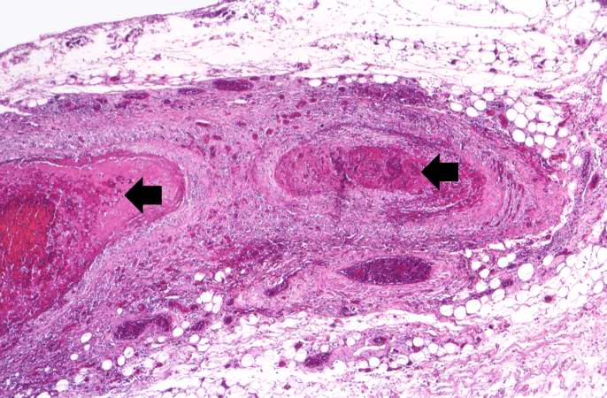
Revision as of 17:56, 20 August 2013 by Peter Anderson (talk | contribs) (This is a higher-power photomicrograph of this mesenteric vessel. Note the thrombotic material occluding the vessel (arrows) and the inflammatory cell infiltrate in the wall of the vessel and in the surrounding adventitia.)
No higher resolution available.
IPLab6PAN5.jpg (686 × 450 pixels, file size: 79 KB, MIME type: image/jpeg)
This is a higher-power photomicrograph of this mesenteric vessel. Note the thrombotic material occluding the vessel (arrows) and the inflammatory cell infiltrate in the wall of the vessel and in the surrounding adventitia.
An infiltrate is an accumulation of cells in the lung parenchyma--this is a sign of pneumonia.
File history
Click on a date/time to view the file as it appeared at that time.
| Date/Time | Thumbnail | Dimensions | User | Comment | |
|---|---|---|---|---|---|
| current | 17:56, 20 August 2013 | | 686 × 450 (79 KB) | Peter Anderson (talk | contribs) | This is a higher-power photomicrograph of this mesenteric vessel. Note the thrombotic material occluding the vessel (arrows) and the inflammatory cell infiltrate in the wall of the vessel and in the surrounding adventitia. |
- You cannot overwrite this file.
File usage
The following page links to this file: