File:IPLab6Hashimoto3.jpg
Revision as of 17:43, 20 August 2013 by Peter Anderson (talk | contribs) (This is a higher-power photomicrograph of thyroid from this case. Note the large number of blue-staining inflammatory cells in this tissue. These cells appear to be forming germinal centers. Some residual thyroid gland tissue can be seen in this sectio...)
No higher resolution available.
IPLab6Hashimoto3.jpg (671 × 450 pixels, file size: 55 KB, MIME type: image/jpeg)
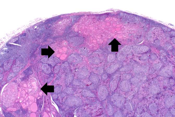
This is a higher-power photomicrograph of thyroid from this case. Note the large number of blue-staining inflammatory cells in this tissue. These cells appear to be forming germinal centers. Some residual thyroid gland tissue can be seen in this section (arrows).
File history
Click on a date/time to view the file as it appeared at that time.
| Date/Time | Thumbnail | Dimensions | User | Comment | |
|---|---|---|---|---|---|
| current | 17:43, 20 August 2013 | | 671 × 450 (55 KB) | Peter Anderson (talk | contribs) | This is a higher-power photomicrograph of thyroid from this case. Note the large number of blue-staining inflammatory cells in this tissue. These cells appear to be forming germinal centers. Some residual thyroid gland tissue can be seen in this sectio... |
- You cannot overwrite this file.
File usage
The following page links to this file: