File:IPLab5Gaucher8.jpg
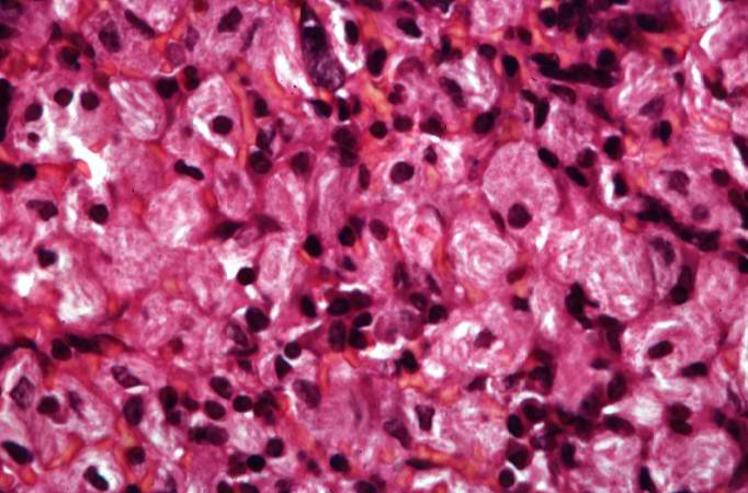
Revision as of 15:12, 20 August 2013 by Peter Anderson (talk | contribs) (This is a higher-power photomicrograph of the spleen from this case. At this higher power individual cells can be better appreciated and the fibrillar nature of the eosinophilic cytoplasmic material can be seen.)
No higher resolution available.
IPLab5Gaucher8.jpg (682 × 450 pixels, file size: 53 KB, MIME type: image/jpeg)
This is a higher-power photomicrograph of the spleen from this case. At this higher power individual cells can be better appreciated and the fibrillar nature of the eosinophilic cytoplasmic material can be seen.
File history
Click on a date/time to view the file as it appeared at that time.
| Date/Time | Thumbnail | Dimensions | User | Comment | |
|---|---|---|---|---|---|
| current | 15:12, 20 August 2013 | | 682 × 450 (53 KB) | Peter Anderson (talk | contribs) | This is a higher-power photomicrograph of the spleen from this case. At this higher power individual cells can be better appreciated and the fibrillar nature of the eosinophilic cytoplasmic material can be seen. |
- You cannot overwrite this file.
File usage
The following page links to this file: