IPLab:Lab 4:Septic Emboli
Clinical Summary
This 4-year-old female sustained second and third degree burns involving approximately forty percent of the body surface. Subsequently, she developed septicemia secondary to skin infection and died in septic shock on the 10th hospital day. Antemortem blood cultures were positive for Enterobacter aerogenes and Staphylococcus aureus.
Images
-
 This gross photograph of lung demonstrates microabscesses due to septic embolization. Note the small 2 to 3-mm lesions scattered throughout this lung tissue (arrows).
 -
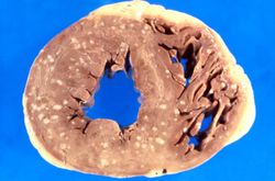 This is a gross photograph of myocardium with multiple embolic lesions scattered throughout the left and right ventricles.
 -
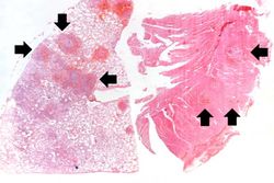 This low-power photomicrograph shows a section of lung on the left and myocardium on the right. Both pieces of tissue have multiple embolic lesions seen as blue staining areas with massive infiltration of inflammatory cells (arrows).
 -
-
-

-
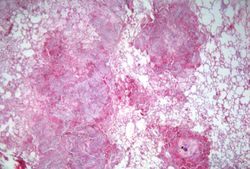
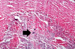 This is a higher-power photomicrograph of the abscess within the myocardium illustrating colonies of bacteria as the dark blue-staining material (arrow).
 -
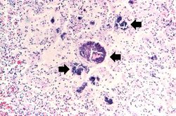
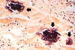 This is a high-powered photomicrograph of a myocardial abscess stained with a special tissue Gram stain (Brown & Brenn) to illustrate the colonies of bacteria in this myocardial tissue (arrows).


Virtual Microscopy
<peir-vm>IPLab4SepticEmboli</peir-vm>
Study Questions
Additional Resources
Reference
- eMedicine Medical Library: Septic Shock
- eMedicine Medical Library: Bacterial Sepsis
- eMedicine Medical Library: Multisystem Organ Failure of Sepsis
- Merck Manual: Sepsis and Septic Shock
- Merck Manual: Bacteremia
Journal Articles
- Hiorns MP, Screaton NJ, Müller NL. Acute lung disease in the immunocompromised host. Radiol Clin North Am 2001 Nov;39(6):1137-51, vi.