File:IPLab3AcuteAppendicitis1.jpg
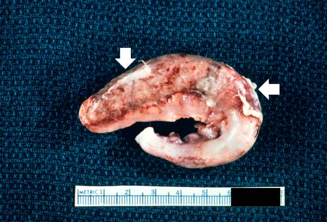
Revision as of 02:10, 19 August 2013 by Seung Park (talk | contribs) (This is a gross photograph of the appendix which was removed from this patient with acute appendicitis. Note the rough, shaggy material (arrows) on the surface due to deposition of fibrin and inflammatory cells.)
No higher resolution available.
IPLab3AcuteAppendicitis1.jpg (660 × 450 pixels, file size: 61 KB, MIME type: image/jpeg)
This is a gross photograph of the appendix which was removed from this patient with acute appendicitis. Note the rough, shaggy material (arrows) on the surface due to deposition of fibrin and inflammatory cells.
File history
Click on a date/time to view the file as it appeared at that time.
| Date/Time | Thumbnail | Dimensions | User | Comment | |
|---|---|---|---|---|---|
| current | 02:10, 19 August 2013 | | 660 × 450 (61 KB) | Seung Park (talk | contribs) | This is a gross photograph of the appendix which was removed from this patient with acute appendicitis. Note the rough, shaggy material (arrows) on the surface due to deposition of fibrin and inflammatory cells. |
- You cannot overwrite this file.
File usage
The following page links to this file: