File:IPLab1MyocardialInfarction3.jpg
Revision as of 19:01, 23 August 2013 by Seung Park (talk | contribs)
No higher resolution available.
IPLab1MyocardialInfarction3.jpg (676 × 450 pixels, file size: 65 KB, MIME type: image/jpeg)
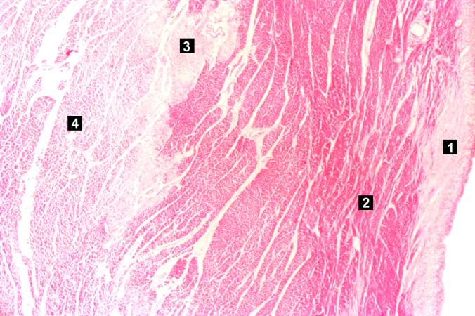
This higher-power photomicrograph shows endocardium on the right side of this image. Directly beneath the endocardium is a pale area consisting of cardiac myocytes exhibiting vacuolar degeneration (1). The area of infarction is visible as a hypereosinophilic area (2) and there is a second zone of vacuolated myocytes (3) between the infarct and the normal myocardium (4).
File history
Click on a date/time to view the file as it appeared at that time.
| Date/Time | Thumbnail | Dimensions | User | Comment | |
|---|---|---|---|---|---|
| current | 13:49, 15 August 2013 | | 676 × 450 (65 KB) | Seung Park (talk | contribs) |
- You cannot overwrite this file.
File usage
The following page links to this file: