IPLab:Lab 11:Schistosomiasis
Images
-
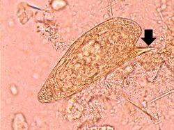 This is a high-power photomicrograph of the patient's fecal specimen containing the characteristic egg of Schistosoma mansoni. Note the spine on the egg (arrow).
 -
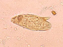 This is high-power photomicrograph of the patient's fecal specimen containing another Schistosoma mansoni egg.
 -
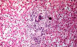 This is a photomicrograph of the liver from another patient with Schistosoma mansoni. These eggs have elicited a robust inflammatory response, including a multinucleated giant cell. These lesions go on to heal by fibrosis resulting in cirrhosis and the classic "pipe-stem" fibrosis.


